
Runner’s Knee (PFPS): What Causes it?
Building off my initial overview post on Runner’s Knee, I will take a deeper dive into different sections related to runner’s knee or patellofemoral pain syndrome (PFPS). In this first deep dive, I will review the pathophysiology or the cause behind PFPS and how it has changed from a pathoanatomical to a more systemic and load related condition. This in-depth guide will give you a better understanding of what's really causing your knee pain and how to better consume the right and most updated ideas on runner’s knee.
Myths of Runner’s Knee
If you have been told you have PFPS solely based on an X ray that shows your kneecap is “sitting wrong” or “tracking funky” then you may want to consider a second opinion. This outdated idea has long been one of the most common explanations for runner’s knee, however we have substantial evidence that shows variations in anatomy do not correlate with those who develop knee pain. Another common fallacy for runner’s knee is related to the VMO or medial thigh muscle. It was said to hold the key to “fixing” kneecap tracking but again we have evidence to show that highly specific VMO exercises and stretching is consistently out performed by hip/knee exercises. A final misnomer for runner’s knee pain is known as chondromalacia. This theory puts the blame on the cartilage on the underside of the kneecap, however we know that articular cartilage is non-innervated or lacks nerves. Again dispelling the idea of a single anatomic factor causing PFPS.
The Multifactorial Shift
Rather than holding to this idea of anatomical causes, the shift in understanding PFPS has transitioned to a multifactorial model. One of the first factors is based on Scott Dye’s work, originally from 1996, related to tissue homeostasis and its “envelope of function.” This means that your tissues have a specific load tolerance (capacity) and when that capacity is exceeded, overload on the tissues can lead to pain. Related to running, this may mean overtraining, rapid increases in mileage or intensity that push the knee beyond its current capacity leading to pain. However, this can be avoided with proper training build up and recovery.
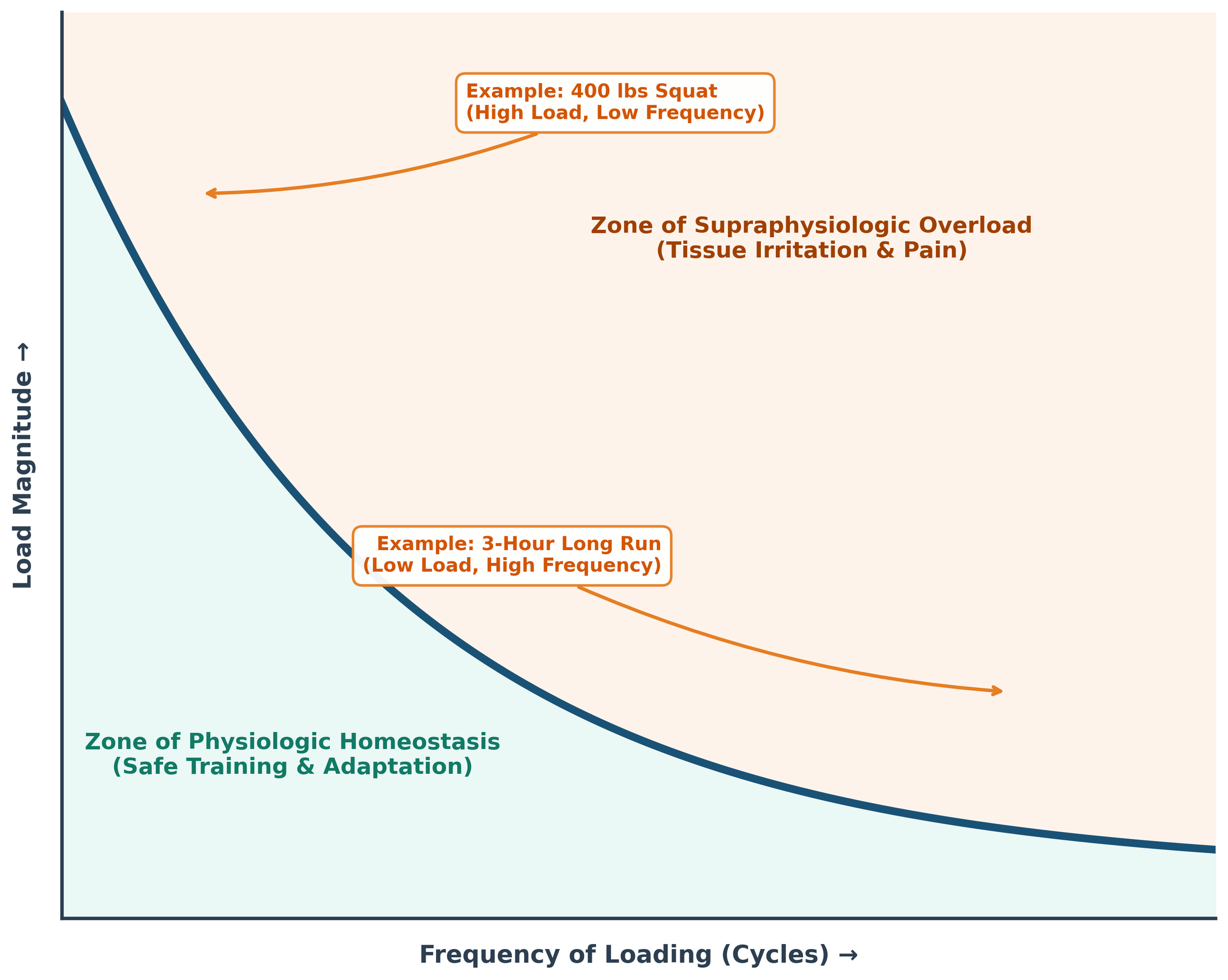
Adapted from Scott Dye’s Envelop of Function Model
The exact tissue cause of pain is still not fully known but there are a few highly innervated areas around the knee joint that may be responsible. These include subchondral bone, synovial membrane, retinaculum, and the Hoffa fat pad, all of which can contribute to symptoms.
Weakness before pain, or pain before weakness?
Another shift related to PFPS is regarding leg strength. Moving away from the idea of isolated VMO strength, studies show that general quad strength is a risk factor in developing PFPS. However at the hip, the story is a little different. Weakness at the hips is not a risk factor for developing PFPS, rather it is a consequence of it. It is thought that due to pain around the knee the brain will down-regulate muscle output or reduce how much the muscles are used around the hip to protect the knee joint. This leads to further development of the dread “hip drop” that is so commonly discussed on social media. In this case, it is often a compensatory strategy utilized by the body in response to the knee pain to avoid deeper knee bends.
You are More than Your Knee
The final part of this multifactorial model of runner’s knee is related to pain, brain, and systemic inflammation. Though runner’s knee may start with local tissue overload and damage, in many cases it shifts to a central nervous system problem. This is called central sensitization as the nervous system becomes hypersensitive to normal and harmless pressures or stimuli which triggers severe pain. Think of fire alarms going off but there is no fire. This is what happens when the knee becomes sensitized.
This then leads into the brain as it develops a kinesiophobia or fear of movement. This fear of movement changes how you move, it forces you to become more stiff, protective, and guarded around the joint. This snowball effect creates higher disability and even more fear around your knee. As you can see, what was once a simple case of knee pain can quickly develop into something much greater.
Your knee, brain, and nervous system are all major components of knee pain but let’s not forget you are a whole person. You have other life stressors like family, a job, bills to pay, on top of training for your next race. These are all important but what is easily forgotten is sleep and systemic inflammation. Poor sleep is directly linked to increased injury risk, reduced pain thresholds, and decreased tissue repair. Add in secondary lifestyle factors like carrying excess weight or having poor nutrition which can further trigger symptoms. This process occurs through the release of pro-inflammatory markers (IL-6 and TNF-alpha) which further break down the joint, increasing inflammation, and sensitizing pathways. This is a biopsychosocial approach. Realizing that you are more than just a knee, you are an ecosystem with many environments interacting with each other.
If you have made it this far, kudos to you as this can be some dense content! I hope that you have found it worth it and learned at least a few things along the way. Remember that, runner’s knee is no longer just a kneecap tracking issue but something much more. It is a multifactorial condition that needs a multifactorial management plan. An approach that I will discuss in the next post to give you the tools to help manage this complex condition.
In the meantime, if you have any questions about PFPS please do not hesitate to reach out via email. And if you are in the CSRA area and are are ready to work with a professional to help manage your knee pain, schedule an appointment today!
Disclaimer: This information is for educational purposes only and does not constitute medical advice, diagnosis, or treatment. Always seek the advice of your physician or other qualified health provider with any questions you may have regarding a medical condition or health-related goals.
References
Dye S. F. (1996). The knee as a biologic transmission with an envelope of function: a theory. Clinical orthopaedics and related research, (325), 10–18. https://doi.org/10.1097/00003086-199604000-00003
Esculier, J.-F., Maggs, K., Maggs, E., & Dubois, B. (2020). A contemporary approach to patellofemoral pain in runners. Journal of Athletic Training, 55(12), 1206–1214. https://doi.org/10.4085/1062-6050-0535.19
Gragnani, B. C., Hart, H. F., Forsyth, A., Barton, C. J., & De Oliveira Silva, D. (2026). Patellofemoral pain is a multifactorial complex condition; are we missing a multidisciplinary approach to its management? Time for a paradigm shift. Open Access Journal of Sports Medicine, 16, 517971. https://doi.org/10.2147/OAJSM.S517971
Neal, B. S., Lack, S. D., Bartholomew, C., & Morrissey, D. (2024). Best practice guide for patellofemoral pain based on synthesis of a systematic review, the patient voice and expert clinical reasoning. British Journal of Sports Medicine, 58(24), 1486–1495. https://doi.org/10.1136/bjsports-2024-108110
Nielsen, R. Ø., Parner, E. T., Nohr, E. A., Lind, M., & Rasmussen, S. (2014). Excessive progression in weekly running distance and risk of running-related injuries: An association which varies according to type of injury. Journal of Orthopaedic & Sports Physical Therapy, 44(10), 739–747. https://doi.org/10.2519/jospt.2014.5164
Rathleff, M. S., Graven-Nielsen, T., Hölmich, P., et al. (2019). Activity modification and load management of adolescents with patellofemoral pain: a prospective intervention study including 151 adolescents. The American Journal of Sports Medicine, 47(7), 1629–1637. https://doi.org/10.1177/0363546519843915
Teng, H.-L., & Powers, C. M. (2014). Sagittal plane trunk posture influences patellofemoral joint stress during running. Journal of Orthopaedic & Sports Physical Therapy, 44(10), 785–792. https://doi.org/10.2519/jospt.2014.5249